Scanning Electron Microscope
The Department of Invertebrate Zoology installed the first Scanning Electron Microscope (SEM), a Zeiss EVO 40XVP, in January 2005. It was purchased with an award from the National Science Foundation (NSF MRI 0420726) and a generous bequest from a private donor. The EVO 40XVP was donated to the Santa Barbara Hackerspace, a non-profit, community tech workshop.
In June 2019, we installed a new instrument with advanced capabilities, a Zeiss EVO 10 LS. The microscope is employed to advance our knowledge and understanding of the natural world, with an emphasis on using the collection resources at SBMNH.
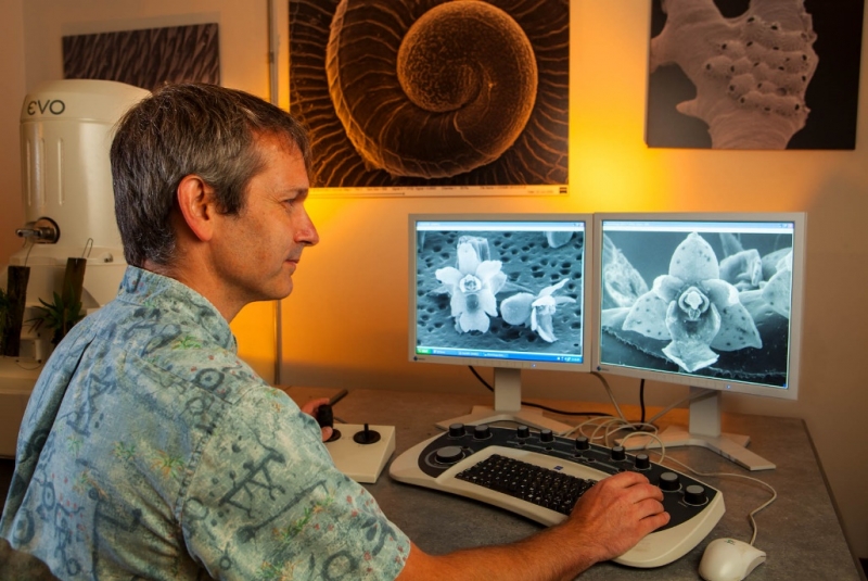
Curator of Malacology Dr. Daniel Geiger uses the SEM in his research on miniature orchids (pictured) and invertebrates, and trains new users of the instrument. Photo by Chuck Place.
Activities
The SEM is used for a variety of activities. Research projects (description of new species, understanding evolutionary history of organisms, morphological and anatomical studies) by in-house researchers, affiliated students, and visiting scholars are the main use. It is also employed in outreach activities, including an exhibit on SEM at SBMNH (including a printed catalog), museum docent training, and SEM demonstrations to the general public, K-12 schools, and students of higher education. Students are also trained to use the instrument.
The Instrument
Zeiss EVO 10 LS
- LaB6 electron source (optional W source), 0.2–30 kV
- Magnification in continuous zoom, 7–1,000,000x
- Resolution to 2 nm under ideal conditions
- Secondary Electron Detector (Everhart-Thornley)
- Zeiss five quadrant High Definition Backscatter Electron Detector
- Zeiss C2DX Extended Range Cascade Current Detector, SE for VP and E-SEM
- Zeiss EDS detector, Zeiss SmartEDX software
- Chamber scope
- Sample camera
- 5 axis motorized stage, compucentric, X = 80 mm, Y = 100 mm, Z = 35 mm, Tilt -10–90˚, Rotation continuous 360˚
- Beam deceleration to -5 kV, HVac only, mixed SE/BSE signal
- Dry N2 venting and VP gas
- VP and E-SEM capable, 10–133 Pa in easy VP, 10–400 Pa in VP, 10–3000 Pa in XVP with beam sleaves
- Water vapor kit
- Deben/Zeiss EVO XVP coolstage -25 to + 50˚C, limited rotation and tilt
- Image size up to 30,000 x 24,000 pixels (~720 MB/MP files 8 bit, 16 bit capable)
- SmartSEM touch: optional easy interface on 23" touchscreen for novice users
- Faraday cup available, for beam current measurement, 50 µm aperture
- Custom 12 mm angled copper stubs for cold stage available
Sputter Coater: Cressington 108E with Rotacota rotating planetary stage, Ar gas, Au target, 12x 12 mm stups
Critical Point Dryer (CPD): Tousimis 815A, 4 and 12 slot sample holders, 100 µm small particle holder
Specimen types
- Hard specimens (shells, insects, rocks): coated (SE and/or BSD detectors), uncoated (VPSE and/or BSD detector)
- Dehydrated soft tissue (HMDS, CPD prepared samples): coated or uncoated
- Hydrated tissues and specimens (coolstage, ESEM)
- Chemical element analysis, mapping, energy dispersive spectroscopy (EDS/EDX/EDAX)
Usage
Training is provided individually to prospective users. Please contact Henry Chaney hchaney@sbnature2.org, 805-682-4711 ext. 150 for details. Time on the SEM is free of charge for internal Museum use.
External users can rent time on the equipment under the following conditions:
- Academic projects: $50/h independent operator, must sign a liability document. $130/h SEM operated by SBMNH personnel. Sputter coater $10 per run, CPD $15/run
- Commercial projects: $120/h independent operator, must sign a liability document. $200/h SEM operated by SBMNH personnel. Sputter coater $15/run, CPD $25/run.
- Minimum duration for session is 1 hour, thereafter billed in 15-minute increments.
- Time for set-up changes from easy VP/HVac, 20 µm intermediate aperture is charged at a higher rate, including aperture changes, beam deceleration holder install, coolstage & ESEM set-up. Take-down time to return instrument back to easy VP/20 µm is also charged at a higher rate.
- For EDS, external user defines/researches analysis conditions (beam current, acceleration voltage) and additionally provides calibration samples, if required. If analysis conditions are unclear, additional research charges apply. Inquire for quote.
New independent users are required to receive training for at least two hours, at $130/h (academic) or $200/h (commercial). Independent user privileges are given at sole discretion of qualified and authorized SBMNH staff. Small amounts of supplies (stubs, mounting media, solvents) are available from SBMNH and are charged to the investigator. For larger amounts, investigators should provide their own supplies. Data is neither stored nor archived at SBMNH. Each investigator or recipient of data is responsible for their own data. Etiological specimens and hazardous materials (e.g., radioactive materials, asbestos) are not permitted in the facility. SBMNH reserves the right to refuse any specimen or proposed procedure. Usage conditions are subject to change at any time and without prior notice.
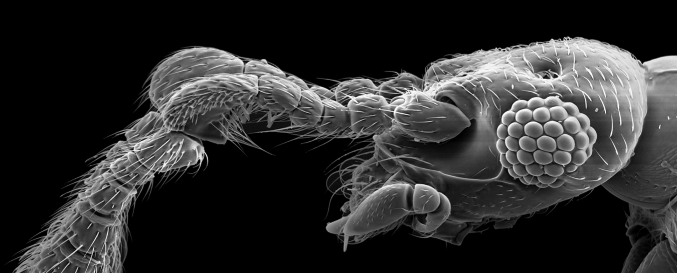
Top image: An undescribed species of Reichenbachia (short-winged mold beetle, Staphylinidae: Pselaphinae), SEM image by Michael Caterino
